Tetralogy of Fallot
 |
| Tetralogy of Fallot |
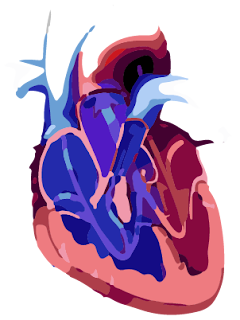
Tetralogy of Fallot is pulmonary stenosis associated with high-mounted ventricular defect. In this way, right ventricle empties into the aorta instead into the pulmonary artery and therefore aortic blood is highly unsaturated with oxygen. Leads to early cyanosis and clubbing of the fingers. Cyanosis is caused by fatigue. This is a problem that exists in the patient since birth. Tetralogy of Fallot occur in just 0.03% of all newborn babies.
The clinical picture of Tetralogy of Fallot
Symptoms and signs of Tetralogy of Fallot
In severe cases of Tetralogy of Fallot a child development is slowed. Dyspnea is a common, squat position facilitates fatigue and dyspnea, and occasionally comes to syncope. Express signs of cyanosis and clubbing of the fingers, moderate distortion of right ventricle and absence of apical pulse, as well as a brief, rough systolic murmur and thrill along the left edge of the sternum. The heart is not enlarged. In a medium-large openings loud second sound can be heard, as he is torn with pulmonary component of reduced amplitude. Because Tetralogy of Fallot causes disturbance of oxygen in the blood, babies have bluish skin, some children never express this symptom if the damage is small.
Depending on the severity of illness of heart, sometimes Tetralogy of Fallot can be detected immediately after birth. Tetralogy of Fallot is a combination four related heart defects: pulmonary stenosis, right ventricular hypertrophy, ventricular septal defect, overriding aorta.
Depending on the severity of illness of heart, sometimes Tetralogy of Fallot can be detected immediately after birth. Tetralogy of Fallot is a combination four related heart defects: pulmonary stenosis, right ventricular hypertrophy, ventricular septal defect, overriding aorta.
Radiographic findings of Tetralogy of Fallot
Lung fields are clear. Apex of the heart is blunt with a concave segment in arteria pulmonalis (heart has shaped of the wooden shoes). Right aortic arch exists in 25% of cases.
ECG of Tetralogy of Fallot
Right ventricular hypertrophy is moderate and almost always present. Occasionally there are also expressed P waves.
Echocardiography of Tetralogy of Fallot
With the help of this test with certainty, we can reveal the presence of Tetralogy of Fallot and we can see the hole between the left and right ventricles or VSD.
Special tests for Tetralogy of Fallot
The diagnosis is made and based on cardiac catheterization and angiocardiography from the right ventricle. In this way and morphological appearance is determined. Aortography is recommended as a routine examination in patients who are in preparation for surgery. With aortography we can show aortic branches and an unexpected associated defects.
Treatment for Tetralogy of Fallot
Tetralogy of Fallot can be treated surgically with the use of the machine for extracorporeal circulation. Operative mortality is moderately low. Patients with underdeveloped pulmonary arteries previously are recommended to Blalock-Taussig type of surgery in which we can increase the flow of blood to the lungs to allow the patient to grow up sufficiently for a complete operation.
Prognosis for Tetralogy of Fallot
In adults the Tetralogy of Fallot is most common cyanotic congenital heart defect, but children do not reach adulthood so often. The most common cause of death is pronounced hypoxemia. Vascular thrombosis caused by polycythemia is also common. Strength of a syndrome is depending on the degree of pulmonary stenosis, if the stenosis is high a left-right shunt and pulmonary blood flow are decreased.
After surgery, patient lead normal live, but he still have to visit the cardiologist to monitor his situation and prescribing appropriate medications. Long term complication after surgery, like right ventricular failure, can appear, and also problem with the heart rate or rhythm. Limited physical activity is recommended. However, if the operation is carried out on time, does not have to be a limitation of physical activity, later in life. Sometimes, doctors recommend antibiotics to the child before dental procedures to prevent infections that can cause endocarditis, inflammation of the the mucous membranes of the heart.
After surgery, patient lead normal live, but he still have to visit the cardiologist to monitor his situation and prescribing appropriate medications. Long term complication after surgery, like right ventricular failure, can appear, and also problem with the heart rate or rhythm. Limited physical activity is recommended. However, if the operation is carried out on time, does not have to be a limitation of physical activity, later in life. Sometimes, doctors recommend antibiotics to the child before dental procedures to prevent infections that can cause endocarditis, inflammation of the the mucous membranes of the heart.